
Intestinal Polyps
Written and medically reviewed by Bahir Hadi, specialist in surgery, PhD
Intestinal Polyps: A Surgical Guide to Types, Risks, and Detection via Colonoscopy
What are Intestinal Polyps?
Intestinal polyps are small growths or nodules that form in the lining of the inside of the large intestine (colon) or rectum. Most polyps are harmless, but certain types, called adenomas, can develop into cancer over time - a process that takes many years.
Some cases of bowel cancer (colorectal cancer) develop from precursor lesions such as adenomas or serrated lesions, but not all polyps turn into cancer. Removing precursor lesions at colonoscopy is a well-documented part of prevention.
Histological Types of Intestinal Polyps
Polyps are classified according to how they look under a microscope (histology). Their type is crucial for their risk of developing into cancer.
1. Precancerous Polyps (Neoplastic)
These polyps have the potential to become malignant (cancerous):
| Type | Description and Malignancy Risk |
|---|---|
| Adenomas | The most common type of precancerous polyp. The main precursor to colorectal cancer among the polyp types found at colonoscopy. |
| Serrated lesions | A group with a saw-toothed growth pattern. The 2019 WHO classification distinguishes hyperplastic polyps (HP), sessile serrated lesions (SSL) with or without dysplasia, and traditional serrated adenomas (TSA). Some SSLs and TSAs can be precursors to bowel cancer; the significance depends among other things on size, dysplasia, number and whether the lesion was completely removed. The serrated pathway differs molecularly from the classic adenoma pathway (WHO 2019 is nomenclature; clinical consequences follow ESGE 2020/BSG). |
2. Harmless Polyps (Non-Neoplastic)
These polyps generally carry no risk of developing into cancer and rarely require further follow-up:
- Hyperplastic Polyps: Part of the serrated group. Small hyperplastic polyps in the rectosigmoid without dysplasia do not normally trigger surveillance on their own (ESGE 2020/BSG); management depends on size, location, number and histology. Telling a hyperplastic polyp from a sessile serrated lesion is a microscopic judgement and can be difficult.
- Inflammatory Polyps: Usually related to inflammatory bowel diseases such as Colitis Ulcerosa.
Dysplasia: The Severity in Adenomas
Dysplasia describes how abnormal the cells in a polyp are. The grade of dysplasia is the most important factor determining the risk that the polyp is already cancerous or will become so.
| Dysplasia Grade | Description | Monitoring/Treatment |
|---|---|---|
| Low-grade dysplasia | The cells are slightly abnormal. The risk of cancer development is low, but the polyp must be completely removed. | Removal during colonoscopy (Polypectomy). |
| High-grade dysplasia | The cells are very abnormal and resemble cancer cells. This is the final step before actual invasive cancer. | Rapid and complete removal, as there is a high risk that the polyp conceals an early stage of cancer. |
Screening Programme in Denmark: Why Colonoscopy is Key
The Danish surveillance programme for intestinal polyps is based on findings made during a colonoscopy (endoscopic examination of the large intestine). The purpose is to remove adenomas and SSLs before they become dangerous.
The follow-up intervals after a polyp has been removed are determined by the doctor (usually a gastroenterologist or surgeon) based on:
- The number of removed polyps.
- The type (histology) of the polyps (e.g., adenoma vs. hyperplastic).
- The grade of dysplasia (low- vs. high-grade).
- The size of the polyps.
| Previous Findings | Recommended Follow-up Interval |
|---|---|
| Low risk (1-4 adenomas under 10 mm with low-grade dysplasia) | Return to the organised bowel cancer screening programme. Where no screening programme applies, the alternative is a repeat colonoscopy after 10 years |
| Increased risk (adenoma ≥10 mm, high-grade dysplasia, ≥5 adenomas, or serrated polyp ≥10 mm or with dysplasia) | 3 years |
| After piecemeal removal of polyps ≥20 mm | Check after 3-6 months, then a further check after about 1 year |
The intervals follow the 2020 guideline from the European Society of Gastrointestinal Endoscopy (ESGE) and assume a complete, good-quality colonoscopy with all polyps fully removed (ESGE 2020, post-polypectomy surveillance).
Two things are easily confused: returning to an organised screening programme is not the same as having a repeat colonoscopy after ten years. The ten-year colonoscopy is what applies where no organised screening is available.
Denmark uses a shared polyp surveillance algorithm from 2023. It specifies return to bowel cancer screening for the relevant low-risk findings, and after piecemeal removal of polyps of at least 20 mm a resection check at six months and a further colonoscopy one year later (Danish polyp surveillance algorithm, 2023).
Important: If you have a family history of bowel cancer or polyps, your individual surveillance programme may differ.
Treatment: Removal of Intestinal Polyps
The goal of treatment is always complete removal of the polyp.
1. Polypectomy during Colonoscopy (Standard Treatment)
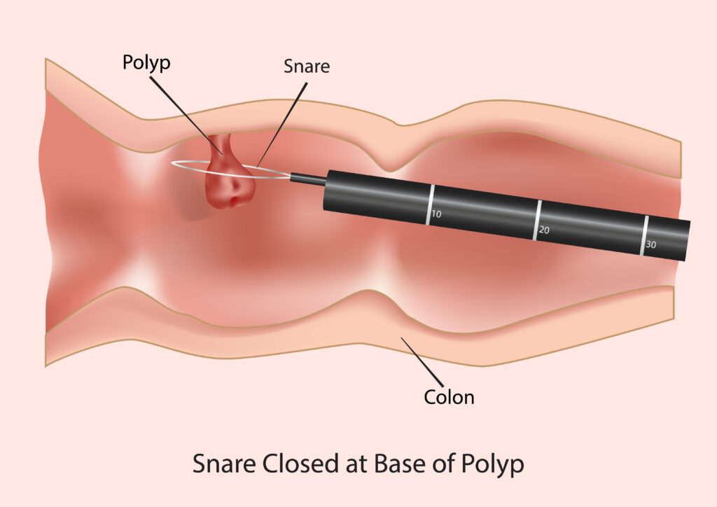
The primary treatment is Polypectomy - removal of the polyp during a colonoscopy.
- Small polyps (up to about 10 mm) are today generally removed with a cold snare, a thin wire loop used without electrical current. In its 2024 update ESGE advises against routine removal with cold biopsy forceps, because more polyp tissue is left behind (ESGE 2024, polypectomy and endoscopic mucosal resection).
- Larger polyps are removed with a snare, often with electrical current and sometimes after injecting fluid beneath the polyp (endoscopic mucosal resection).
- Early Cancer in a Polyp: If a polyp is removed completely and the histology shows an early stage of cancer without deep invasion, removal during the colonoscopy may be the only necessary treatment (curative).
2. Surgical Removal
Surgery does not automatically follow from a large polyp, from high-grade dysplasia or from an incomplete removal. The first question is whether the polyp tissue can be removed completely by endoscopy, if necessary at a unit experienced in advanced polyp removal. High-grade dysplasia is a change within the polyp and does not in itself mean the bowel must be operated on.
Surgery may become relevant if:
- The polyp tissue cannot be removed completely by endoscopy, including after referral for advanced endoscopic treatment.
- Invasive cancer is present with features suggesting a risk of lymph node spread. The operation is then assessed separately in a multidisciplinary setting, weighing benefit against the risk of bowel surgery.
- Residual or recurrent polyp tissue persists despite repeated endoscopic treatment.
Conclusion: Prevention Through Colonoscopy
Intestinal polyps are a common but manageable condition, frequently seen during colonoscopy. Knowledge of histological types, especially adenomas and serrated polyps, and their risk of dysplasia is essential.
At Kirurgen.dk, we emphasise correct investigation and treatment, where colonoscopy plays the most vital role - both in diagnosis and in the preventive removal of potentially dangerous growths.
If you have symptoms, belong to a risk group, or simply wish to understand more about colonoscopy and polyps, we are ready to advise you.
Reference List (PubMed-based Sources)
General Background and Prevention
Intestinal Polyps as a Precursor to Cancer (The Adenoma-Carcinoma Sequence):
Source: Leslie, A., Carey, F. A., Pratt, N. R., & Steele, R. J. (2002). The adenoma-carcinoma sequence. British Journal of Surgery, 89(7), 842-852.
Supports: The claim that bowel cancer almost always arises from a polyp (adenoma) and that removal prevents cancer.
The Effect of Colonoscopy and Polypectomy on the Incidence of Colorectal Cancer:
Source: Zauber, A. G., Winawer, S. J., O'Brien, M. J., Lansdorp-Vogelaar, I., van Ballegooijen, M., Hankey, B. F., Shike, M., Davies, R. J., Fromkes, J. J., Kewenter, J., & Waye, J. D. (2012). Colonoscopic polypectomy and long-term prevention of colorectal-cancer deaths. The New England Journal of Medicine, 366(8), 687-696.
Supports: The importance of colonoscopy as a preventive method and that polyp removal reduces mortality from colorectal cancer.
Histological Types and Risks
Classification and Risk of Serrated Polyps (Sessile Serrated Lesions - SSL):
Source: Bettington, M., Walker, N., Rosty, C., Brown, I., Clouston, A., Goh, R., Weninger, M., Embasse, N., Kana, R., & Appleyard, M. (2016). Sessile serrated adenomas (polyps) represent a pathway for colorectal cancer development. Gut, 65(2), 333-342.
Supports: The classification of precancerous polyps and the inclusion of serrated polyps/SSL in the group with malignant potential.
Dysplasia (Low- and High-Grade) as a Risk Factor:
Source: Dekker, E., Rex, D. K., Anastassiades, G., Arebi, N., Bull, P., Ciaccio, E. J., Clark, M., Colonoscopy Quality Improvement Initiative (CQII) International Expert Group, East, J. E., Farhat, S., Fasoli, M., Fini, L., Halligan, S., Höglund, P., Hurlstone, P., Jover, R., Kaminski, M. F., Kudo, S. E., Løberg, M.,... Gschmeidler, H. (2015). Colorectal Polyp and Cancer Surveillance; Clinical guidelines and quality measures. United European Gastroenterology Journal, 3(6), 493-503.
Supports: The importance of dysplasia (especially high-grade dysplasia) as the most significant factor for cancer risk and as a guide for treatment strategy.
Monitoring and Treatment
Principles for Follow-up Intervals After Polypectomy:
Source: European Society of Gastrointestinal Endoscopy (ESGE) guideline for post-polypectomy surveillance: update 2020.
Source (related): Hassan, C., Quintero, E., Dumonceau, J. M., Regula, J., Brandao, C., Chaussade, S., Dekker, E., Dinis-Ribeiro, M., Ferlitsch, M., Freitas, D., Gralnek, I., Jover, R., Kalager, M., Loberg, M., Machado, J., Mazzotti, F., Repici, A., Rubenstein, J., Simon, H. W., & Stoffel, E. (2020). Post-polypectomy colonoscopy surveillance: European Society of Gastrointestinal Endoscopy (ESGE) guideline-update 2020. Endoscopy, 52(9), 687-707.
Supports: The clinical guidelines forming the basis for the mentioned surveillance intervals (1, 3, 5+ years) based on the number, type, and size of polyps.
Surgical Treatment vs. Endoscopic Removal of Early Cancer in a Polyp:
Source: Sunkara, T., & Gaduputi, V. (2018). Endoscopic treatment of colorectal polyps with early cancer. World Journal of Gastroenterology, 24(25), 2686-2697.
Supports: The criteria for when a polyp containing early cancer can be treated curatively endoscopically (polypectomy) versus when surgical resection is required.
More on this topic at Kirurgen.dk
Category: Endoscopy