
Diverticulitis vs. diverticulosis
Written and medically reviewed by Bahir Hadi, specialist in surgery, PhD
Published: 21 July 2026
Diverticulitis vs. Diverticulosis: What's the difference, and when is it dangerous?
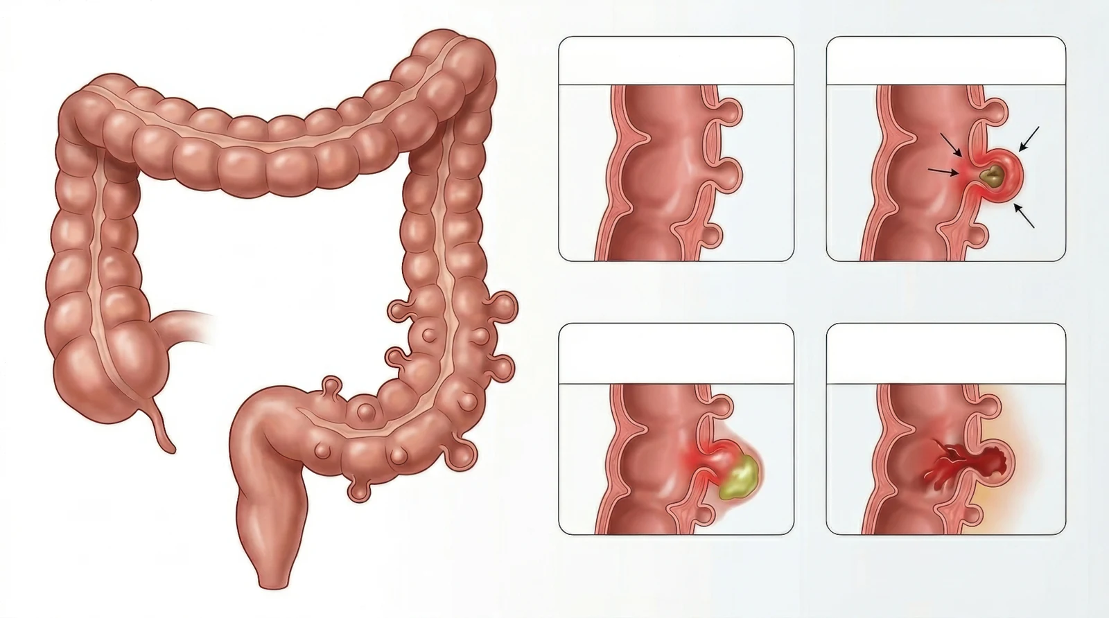
Many people confuse diverticulosis and diverticulitis - but the difference is crucial. The first is a common, usually harmless condition. The second is an acute inflammation that may require hospital admission or surgery.
Diverticulosis: Small pouches in the large intestine
Diverticula are small pouches (typically 5-10 mm) in the bowel wall, which form at weak points where blood vessels penetrate the muscle layer. They are predominantly found in the sigmoid colon (the S-shaped part of the large intestine).
The prevalence rises clearly with age. Diverticula are unusual before the age of 40 and become common in older people; United States surveys describe more than half of people over 60 as having diverticula [1]. These figures come from American data, not Danish registers. Most are discovered incidentally during a colonoscopy or CT scan.
Risk factors include:
- Low fibre intake and a Western diet [2]
- Obesity and physical inactivity [3]
- Smoking and NSAID use [4]
- Genetic predisposition. In a Swedish twin study, heritable factors explained around 40% of the variation in risk at population level, not in the individual [5]
Read also our article on diverticulosis.
Diverticulitis: When a pouch becomes inflamed
Older estimates of a 10-25% lifetime risk after diverticulosis is found are now considered too high. In a retrospective US VA study of patients with incidentally found diverticulosis, 4.3% developed diverticulitis over an average of 11 years of follow-up using a broad definition, while 1% met a strict definition requiring imaging or operative confirmation [6]. The results cannot simply be transferred to all patient groups. Diverticulitis is an acute inflammation or micro-perforation of a diverticulum, and the condition is divided into:
- Uncomplicated diverticulitis (the large majority of cases): Localised inflammation without an abscess, perforation, or fistula
- Complicated diverticulitis (around 20% of cases in published series): Abscess, free perforation, obstruction, or fistula
Symptoms
- Persistent pain in the lower left part of the abdomen ("left-sided appendicitis")
- Fever and elevated CRP
- Change in bowel habits (constipation or diarrhoea)
- Nausea, bloating
- With complications: severe pain, signs of peritonitis, urinary symptoms (fistula to the bladder)
The diagnosis is confirmed with a CT scan of the abdomen, which also classifies the severity using the Hinchey classification (originally described by Hinchey et al. [7], now applied in a CT-based form according to international surgical guidelines [11]).
Treatment of uncomplicated diverticulitis
Previously, almost all cases were treated with antibiotics and hospital admission. Modern Danish and European guidelines now recommend:
- Outpatient treatment without antibiotics in selected patients (immunocompetent, without complications, CRP <140, no fever) [8]
- Rest, clear fluids gradually progressing to a normal diet
- Pain relief (paracetamol; avoid NSAIDs)
The landmark AVOD study (Sweden) and the Dutch DIABOLO study have documented that antibiotics do not speed up recovery or reduce complications in uncomplicated diverticulitis [9,10].
Treatment of complicated diverticulitis
- Smaller abscess: Antibiotics alone are often sufficient
- Larger abscess: Percutaneous drainage combined with antibiotics where drainage is technically feasible; otherwise antibiotics alone initially, if the clinical condition permits
WSES uses 4 cm as the cut-off in its CT classification, but size is not an isolated treatment rule. The choice also depends on where the abscess lies, whether it can be reached for drainage, and how unwell the patient is. The decision rests with the treating surgical department [11]
- Perforated diverticulitis with diffuse peritonitis: In perforated diverticulitis with diffuse peritonitis the surgical method depends on haemodynamic stability, comorbidity and the operative findings. WSES recommends a Hartmann's procedure in critically ill patients and in selected patients with significant comorbidity. In clinically stable patients without significant comorbidity, primary resection with anastomosis, with or without a diverting stoma, may be considered. The specific method is decided during the acute surgical care [11]
- Free air without diffuse peritonitis: Free air on CT without diffuse intra-abdominal fluid or peritonitis does not automatically mean the same treatment. In carefully selected, clinically stable patients, non-operative management with close monitoring may be possible [11]
- Recurrent or complicated cases: A planned elective sigmoid resection is considered - but NOT routinely after 2 attacks as was previously the case; the decision is made on an individual basis [11, 12]
Prevention
- Fibre in the diet: In the EPIC-Oxford cohort of 47,033 UK adults followed for a mean of 11.6 years, participants in the highest fifth of fibre intake had fewer hospital admissions or deaths from diverticular disease than those in the lowest fifth [13]. This is an observed association in a cohort of health-conscious participants, not proof that fibre prevents the disease, and no gram target is given here. How much fibre suits you depends on your symptoms and your other treatment
- Physical activity and a normal weight
- Avoid smoking and frequent NSAID use
- It was previously recommended to avoid nuts, seeds, and popcorn - this has now been disproven and is no longer advised against [14]
When should you contact a doctor?
Seek medical advice for:
- Persistent pain in the lower left abdomen for more than 24 hours
- Fever accompanied by abdominal pain
- Severe bleeding from the rectum
- Signs of peritonitis (rigid abdomen, severe pain, feeling generally unwell) → call 112
References
- Peery AF, et al. Burden and cost of gastrointestinal, liver, and pancreatic diseases in the United States. Gastroenterology 2019;156(1):254-72.
- Aune D, et al. Dietary fibre intake and the risk of diverticular disease: a systematic review and meta-analysis. Eur J Nutr 2020;59(2):421-32.
- Strate LL, et al. Obesity increases the risks of diverticulitis and diverticular bleeding. Gastroenterology 2009;136(1):115-22.
- Humes DJ, et al. Smoking and the risk of diverticular disease. Aliment Pharmacol Ther 2011;34(8):988-94.
- Granlund J, et al. The genetic influence on diverticular disease - a twin study. Aliment Pharmacol Ther 2012;35(9):1103-7.
- Shahedi K, et al. Long-term risk of acute diverticulitis among patients with incidental diverticulosis. Clin Gastroenterol Hepatol 2013;11(12):1609-13.
- Hinchey EJ, et al. Treatment of perforated diverticular disease of the colon. Adv Surg 1978;12:85-109.
- Schultz JK, et al. European Society of Coloproctology: guidelines for the management of diverticular disease of the colon. Colorectal Dis 2020;22(Suppl 2):5-28.
- Chabok A, et al. Randomized clinical trial of antibiotics in acute uncomplicated diverticulitis (AVOD). Br J Surg 2012;99(4):532-9.
- Daniels L, et al. Randomized clinical trial of observational versus antibiotic treatment for a first episode of CT-proven uncomplicated acute diverticulitis (DIABOLO). Br J Surg 2017;104(1):52-61.
- Sartelli M, et al. 2020 update of the WSES guidelines for the management of acute colonic diverticulitis in the emergency setting. World J Emerg Surg 2020;15:32.
- Hall J, et al. The American Society of Colon and Rectal Surgeons Clinical Practice Guidelines for the Treatment of Left-Sided Colonic Diverticulitis. Dis Colon Rectum 2020;63(6):728-47.
- Crowe FL, et al. Diet and risk of diverticular disease in Oxford cohort of EPIC. BMJ 2011;343:d4131.
- Strate LL, et al. Nut, corn, and popcorn consumption and the incidence of diverticular disease. JAMA 2008;300(8):907-14.
Read also
More on this topic at Kirurgen.dk
Category: Gastrointestinal