
Venous leg ulcer
Written and medically reviewed by Bahir Hadi, specialist in surgery, PhD
Venous leg ulcer (ulcus cruris venosum): Causes and modern treatment
A venous leg ulcer - ulcus cruris venosum - is the most severe stage of chronic venous insufficiency. In UK population surveys, active (open) leg ulcers of any cause are found in around 1.5-3 per 1,000 people, rising to about 20 per 1,000 among people over 80 [1]. These figures cover all active leg ulcers, not only those with a venous cause, and they say nothing about an individual's risk in Denmark. The ulcers heal slowly, recur frequently and are often the result of many years of untreated varicose veins. The good news: in the randomised EVRA trial, which included patients with venous leg ulcers of less than six months' duration and superficial venous reflux suitable for endovenous treatment, ulcers healed faster when compression was combined with early endovenous ablation within two weeks, compared with compression where endovenous treatment was deferred until after ulcer healing, or until six months after randomisation if the ulcer had not yet healed. Median healing time was 56 days versus 82 days. Both groups received compression throughout [2].
What is a venous ulcer?
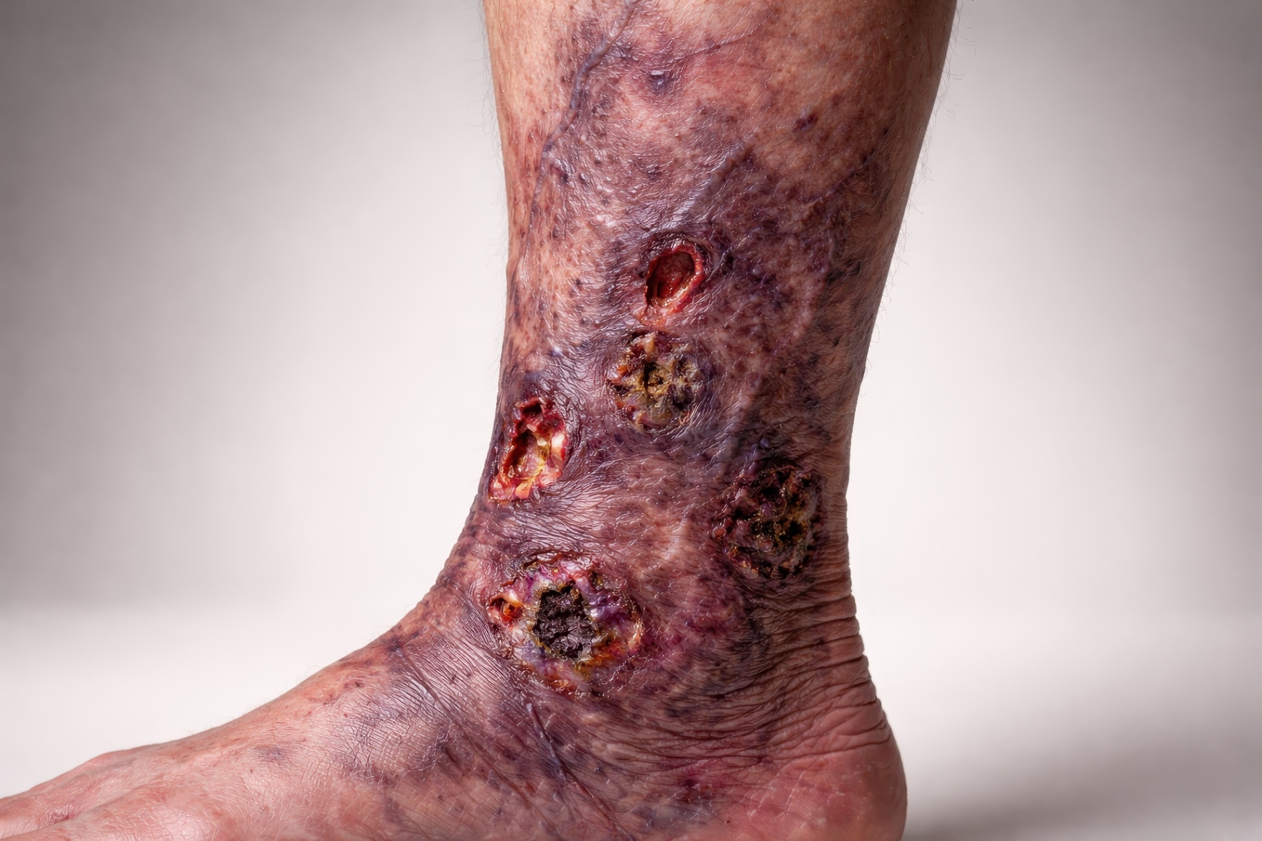
A venous ulcer is a chronic skin defect - typically on the medial ankle ("gaiter area") - that does not heal within 4-6 weeks. It develops when chronic venous hypertension damages the skin and subcutaneous tissue:
- Defective venous valves → blood flows backwards (reflux)
- Elevated venous pressure in the lower leg
- Leakage of red blood cells and protein through the capillary wall
- Inflammation, fibrosis and oxygen deficiency in the skin
- The skin barrier breaks down → ulcer
The stages before a venous ulcer (CEAP)
Venous disease is classified by CEAP [3]:
| Stage | Signs |
|---|---|
| C0-C1 | No visible signs / spider veins |
| C2 | Visible varicose veins |
| C3 | Swollen legs and ankles |
| C4a | Pigmentation, venous eczema |
| C4b | Lipodermatosclerosis, atrophie blanche |
| C5 | Healed venous ulcer |
| C6 | Active venous ulcer |
By the time you reach C4-C5, the risk of a full venous ulcer is markedly increased - and at this stage treatment of varicose veins has the greatest preventive value.
Symptoms and characteristics
- Ulcer on the inside of the ankle, rarely on the foot or above the knee
- Irregular edges, shallow base, yellow-red wound tissue
- Surrounding brown pigmentation and venous eczema
- Chronic swelling that improves with elevation
- Pain (often less than with arterial ulcers)
- Odour and exudate if infected
When to see a doctor?
Seek medical attention immediately if you have:
- An ulcer on the leg that does not heal within 2 weeks
- Increasing pain, redness or fever (infection)
- Sudden worsening of a chronic ulcer
- Ulcer + diabetes or peripheral arterial disease (different treatment required)
Among selected patients with relatively recent venous leg ulcers (less than six months' duration) and superficial venous reflux suitable for treatment, early endovenous ablation together with compression gave faster ulcer healing than deferred treatment, and long-term follow-up showed more ulcer-free time [2,7]. Both groups received compression. The result cannot simply be applied to all chronic leg ulcers, and there is no single figure for how much the risk of a chronic ulcer falls.
Work-up
- Clinical examination - CEAP classification, ulcer size, location.
- Duplex ultrasound (duplex scan) - used to locate valve failure and reflux and to plan treatment in relevant patients [3].
- Ankle-brachial index (ABI) - a pressure measurement used to assess whether arterial supply to the leg is also reduced. It matters before compression. The reading can be falsely high when the arteries are stiff and calcified, for example in diabetes or kidney disease, so a reassuring value does not reliably exclude arterial disease. Where suspicion remains, toe pressure or ultrasound is added (Lægehåndbogen on blood pressure measurement in the lower limbs).
- Wound culture if signs of infection.
- Biopsy for ulcers >3 months without healing (to exclude malignancy).
Modern treatment
1. Compression - the cornerstone [5]:
- Class III compression stocking (34-46 mmHg) or multilayer bandage
- Strong compression requires an ankle-brachial index above 0.8. That is a threshold for how much compression can be applied, not the same as normal arterial circulation
- Correctly applied compression heals a large share of ulcers over some months [5]
2. Endovenous laser ablation (EVLA/EVLT):
The randomised EVRA trial (NEJM 2018) compared compression combined with early endovenous ablation within two weeks against compression where endovenous treatment was deferred until after ulcer healing, or until six months after randomisation if the ulcer had not yet healed:
- Median healing time 56 days versus 82 days. This is not a comparison with compression alone as permanent treatment, but with deferred endovenous treatment
- In the extended follow-up, where participants were followed for up to five years and median follow-up was about 3.5 years (roughly 1,286 days), the overall burden of recurrent ulcers was lower after early treatment. However, there was no statistically certain difference in time to first recurrence (HR 0.82; 95% CI 0.57-1.17). The recurrence rate was 0.11 versus 0.16 per person-year. Cost-effectiveness was calculated over three years, and the authors describe results beyond four years as uncertain [7]
- Early ablation of incompetent truncal veins is a recommendation in international venous ulcer guidelines [2,6]
The treatment is performed under local anaesthesia. How long it takes depends on how many veins are treated and how long they are.
3. Wound care:
- Atraumatic dressings (foam, hydrofibre, alginate by wound type)
- Local debridement, possibly larval therapy for necrosis
- Antibiotics only for clinical infection - not for colonisation
4. Prevention of recurrence:
- Lifelong compression stocking (class II)
- Weight loss if overweight
- Daily exercise → activates the calf muscle pump
- Skin care with moisturiser
Treatment at Kirurgen.dk
We offer Doppler ultrasound and endovenous laser ablation (EVLA/EVLT) after a GP referral. We see patients with both active and healed venous ulcers. Referral and payment are explained on our prices page. See also our articles on treatment of varicose veins, heavy, tired legs and swollen legs and ankles.
References
- Nelson EA, Adderley U. Venous leg ulcers. BMJ Clin Evid 2016;2016:1902.
- Gohel MS, Heatley F, Liu X, et al. A randomized trial of early endovenous ablation in venous ulceration (EVRA). N Engl J Med 2018;378(22):2105-14.
- Lurie F, et al. The 2020 update of the CEAP classification system. J Vasc Surg Venous Lymphat Disord 2020;8(3):342-52.
- O'Donnell TF Jr, et al. Management of venous leg ulcers: clinical practice guidelines of the SVS and AVF. J Vasc Surg 2014;60(2 Suppl):3S-59S.
- O'Meara S, Cullum N, Nelson EA, Dumville JC. Compression for venous leg ulcers. Cochrane Database Syst Rev 2012;(11):CD000265.
- Gloviczki P, Lawrence PF, Wasan SM, et al. The 2023 SVS/AVF/AVLS clinical practice guidelines for the management of varicose veins of the lower extremities. Part II. J Vasc Surg Venous Lymphat Disord 2024;12(1):101670. DOI: 10.1016/j.jvsv.2023.08.011. PubMed
- Gohel MS, Mora MSc J, Szigeti M, et al. Long-term clinical and cost-effectiveness of early endovenous ablation in venous ulceration: a randomized clinical trial (EVRA, extended follow-up up to 5 years). JAMA Surg 2020;155(12):1113-21. PubMed
More on this topic at Kirurgen.dk
Category: Varicose veins