
Barrett's oesophagus
Written and medically reviewed by Bahir Hadi, specialist in surgery, PhD
Published: 15 August 2026
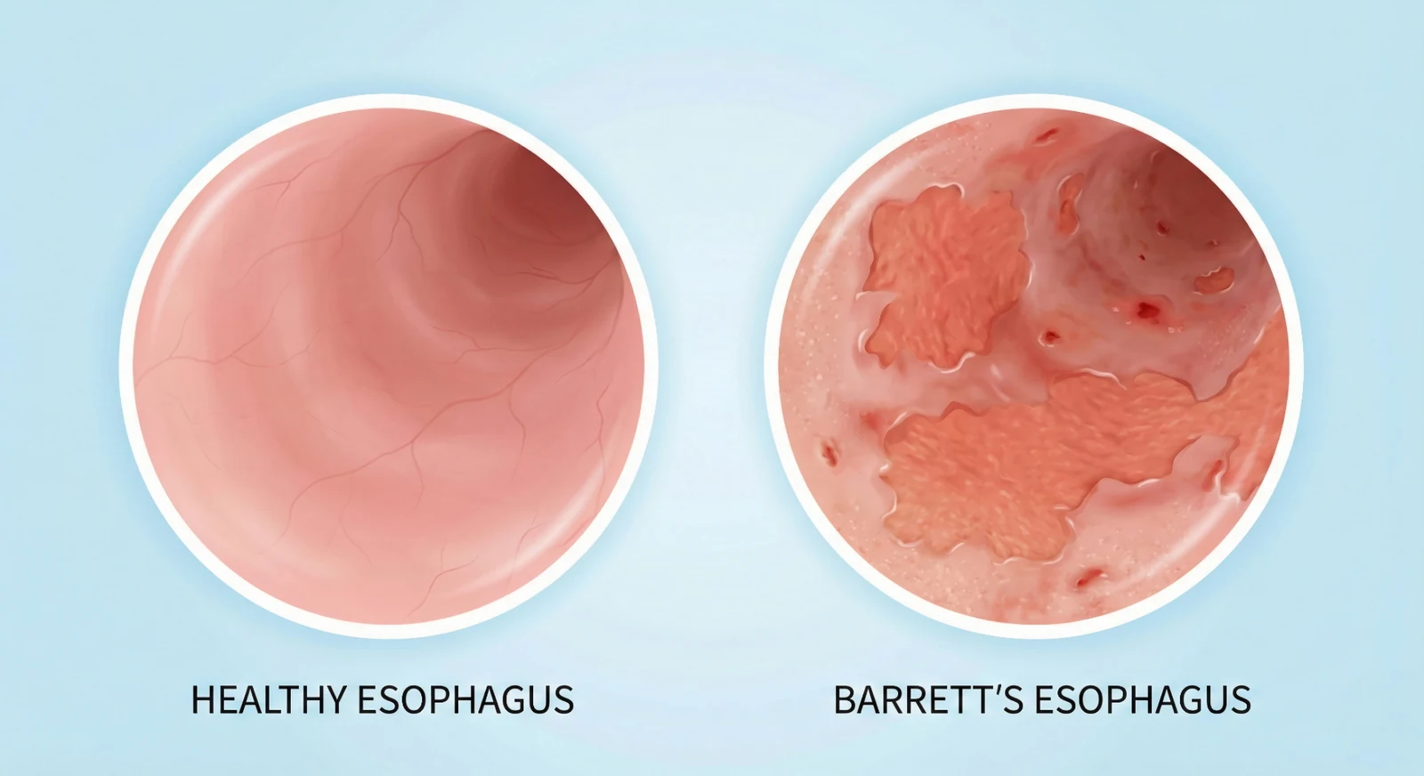
Barrett's oesophagus is a condition where the normal multi-layered squamous cell lining in the lower part of the oesophagus is replaced by a glandular lining (intestinal type, intestinal metaplasia). This change is a consequence of chronic acid exposure - typically after many years of gastro-oesophageal reflux disease (GORD) - and is the most important known risk factor for developing adenocarcinoma of the oesophagus.
The actual risk of developing cancer is low. In a nationwide Danish cohort of 11,028 patients with newly diagnosed Barrett's oesophagus followed from 1992 to 2009, the annual risk of oesophageal adenocarcinoma was 0.12% (95% CI 0.09-0.15) [6]. That figure is the estimate from that specific cohort and patient population, not a universal risk for everyone with Barrett's oesophagus, but the risk is real, and therefore Denmark has well-defined recommendations for investigation, follow-up, and treatment. This article describes how we at Kirurgen.dk manage Barrett's oesophagus. Each recommendation is tied to its own source along the way: the Danish DSGH treatment guideline, last revised in 2016 [1], and the 2023 ESGE guideline [2]. Where we mention British practice, we refer to NICE NG231 [7], which together with ESGE 2023 has superseded the older BSG guideline [3].
What is Barrett's oesophagus?
The lining of the oesophagus is normally pink and covered by squamous cells. In Barrett's oesophagus, a salmon-coloured, velvety lining is seen instead, growing upwards from the junction between the oesophagus and the stomach (the Z-line). The definitive diagnosis is made by endoscopy + biopsy:
- Endoscopically: a visible columnar lining that extends ≥ 1 cm proximally from the gastro-oesophageal junction.
- Histologically: demonstration of specialised intestinal metaplasia with goblet cells.
Both criteria must be met before the diagnosis of "Barrett's oesophagus" is made [1,3].
Prague C&M Classification
The extent is described using the Prague C&M classification, where:
- C = the circumferential (all-round) length of the metaplastic segment in cm.
- M = the maximum length measured from the Z-line to the highest tongue of metaplasia in cm.
For example, C2M5 means that the bottom 2 cm are covered all around by Barrett's lining, and that the longest tongue extends up to 5 cm. The classification is crucial for determining how often the patient requires follow-up.
Who is at risk?
Barrett's oesophagus is most commonly seen in adults with several of the following risk factors [1,3]:
- Chronic reflux symptoms for ≥ 5 years (heartburn, acid regurgitation)
- Age ≥ 50 years
- Male sex
- White (Caucasian) ethnicity
- Central obesity (apple shape)
- Current or former smoker
- First-degree relative with Barrett's oesophagus or oesophageal adenocarcinoma
The Danish guideline states that patients with several of the risk factors above may be offered a gastroscopy, but stresses that screening has not shown a definite effect on cancer survival and is therefore not directly recommended in international guidelines [1]. General population screening is not recommended.
Symptoms - and why the diagnosis is often incidental
Barrett's oesophagus does not cause any specific symptoms in itself. Most patients are referred for a gastroscopy because of typical reflux symptoms:
- Heartburn behind the breastbone
- Acid regurgitation
- Difficulty swallowing (dysphagia)
- Chronic cough, hoarseness or asthma-like problems
"Red flag" symptoms that should trigger an urgent gastroscopy include [1,3]:
- Difficulty or pain on swallowing
- Unexplained weight loss
- Vomiting blood or black stools (see also our article on black stools (melaena))
- Chronic anaemia with no other explanation
- Persistent vomiting
Investigation - The Seattle Protocol
Once a Barrett's lining has been identified at endoscopy, the biopsy strategy is crucial to avoid missing dysplasia or early cancer.
The Danish guideline and ESGE recommend the Seattle biopsy protocol [1,2]:
- Targeted biopsies from all visible irregularities (nodules, ulcers, colour changes). Use the Paris classification to describe their shape and height.
- 4-quadrant biopsies every 2 cm throughout the entire Barrett's segment (every 1 cm if dysplasia is known).
- Biopsies are sent in separate, labelled pots according to their level.
- The use of high-definition endoscopy (HD-WLE) and virtual chromoendoscopy (NBI or similar) increases the detection of dysplasia and should be standard practice [1,2].
The histology is assessed by a pathologist with a subspeciality in gastroenterology, and a finding of dysplasia must always be confirmed by a second pathologist before treatment is initiated [1,3].
Follow-up - depends on segment length and dysplasia
The follow-up below follows the Danish clinical guideline from DMCG/DECG, version 3.0, professionally approved in January 2025 [1]. The Danish pathway starts with a repeat gastroscopy after 6 months of double-dose PPI, to allow for the first biopsies having missed the relevant area; only then is the further interval set. The 2023 ESGE guideline [2] uses the same principle of segment length and dysplasia but with slightly different cut-offs: no surveillance for a segment under 1 cm, every 5 years for 1 to under 3 cm, every 3 years for 3 to under 10 cm, and referral to an expert centre at 10 cm or more. The specific plan is always set by the treating department. The main lines of the Danish programme are:
| Finding | Recommended follow-up |
|---|---|
| Columnar epithelium without goblet cells, no dysplasia | Repeat gastroscopy after 6 months of double-dose PPI; if goblet cells are still absent, surveillance can be stopped |
| Intestinal metaplasia with goblet cells, no dysplasia | Repeat gastroscopy after 6 months of double-dose PPI; then surveillance every 3 years for a long segment (≥ 3 cm) and every 5 years for a short segment (< 3 cm) |
| Patients over 75 | Continued surveillance is assessed individually |
| Epithelial change, indefinite for dysplasia or reactive | Repeat gastroscopy after 6 months of double-dose PPI; review by at least two pathologists with upper GI expertise |
| Low-grade dysplasia (LGD) confirmed by two pathologists | Repeat gastroscopy after 6 months; endoscopic treatment (ablation or EMR) at a highly specialised unit, then surveillance after 6 months and again at 1, 3 and 5 years |
| High-grade dysplasia (HGD) or early cancer | Endoscopic resection (EMR/ESD) at a highly specialised unit; surveillance after 3 and 6 months and again at 1, 2, 3, 4, 5, 7 and 10 years |
The Danish guideline requires that all dysplasia and all unresolved epithelial changes are reviewed by at least two pathologists with upper gastrointestinal expertise, and that dysplastic Barrett's oesophagus is treated and followed at a highly specialised unit [1].
Treatment
1. Optimal Acid Suppression
All patients with Barrett's oesophagus must be treated with a proton pump inhibitor (PPI) in a sufficient dose to control reflux symptoms and keep the lining free of visible oesophagitis [1,3]. PPIs probably reduce - but do not eliminate - the risk of progression to dysplasia.
Read more about acid suppression and lifestyle in our article on GORD / acid reflux.
2. Lifestyle Changes
The same measures as for GORD are recommended:
- Weight loss if overweight
- Smoking cessation
- Elevating the head of the bed and no meals in the last 2-3 hours before bedtime
- Reducing alcohol, coffee and known triggers
3. Endoscopic treatment of dysplasia and early cancer
When dysplasia or intramucosal carcinoma is detected, treatment today is endoscopic and takes place at expert centres [1,2,3]:
- Endoscopic mucosal resection (EMR) or endoscopic submucosal dissection (ESD) of visible lesions - both for diagnosis and treatment.
- Radiofrequency ablation (RFA) of the remaining Barrett's segment to remove the risk of new dysplastic foci.
- Cryotherapy may be used as an alternative to RFA.
For high-grade dysplasia and T1a adenocarcinoma, international guidelines recommend endoscopic treatment rather than oesophagectomy in suitable patients. The recommendation rests on observational studies and guideline appraisal, not on randomised comparisons of the two strategies. The outcome depends on how far the change extends and whether it can be removed completely [2,3].
4. Anti-reflux Surgery
Anti-reflux surgery (typically laparoscopic fundoplication) is not routinely recommended for cancer prevention in Barrett's oesophagus [1,3]. The procedure may be considered for patients with persistent reflux symptoms despite optimal medical treatment, or for volume reflux that PPIs cannot manage.
When should you be referred?
As a patient, you should talk to your GP about a referral for gastroscopy if you have:
- Reflux symptoms for more than 5 years along with several risk factors (age, male sex, obesity, smoking, family history).
- "Red flag" symptoms as described above - regardless of age.
- A previous finding of Barrett's oesophagus without a follow-up plan.
At Kirurgen.dk, we perform oral gastroscopy and nasal gastroscopy (see nasal vs. oral gastroscopy). Which method is relevant, and how the visit is arranged, is agreed individually. Questions about referral, cover and price are clarified with the clinic or your GP before treatment; see contact.
Prognosis
For the individual patient with Barrett's oesophagus without dysplasia, the annual risk of developing adenocarcinoma is low. The absolute annual risk of adenocarcinoma was 0.12% (95% CI 0.09-0.15) in a nationwide Danish cohort of 11,028 patients with a median follow-up of 5.2 years. The estimate applies at group level and cannot be used as an individual prognosis. The risk is higher with dysplasia. 0.12% corresponds to 1.2 cases per 1,000 person-years, and the incidence estimate was calculated after excluding cancers diagnosed within the first year [6]. The estimate must not be transferred to confirmed low- or high-grade dysplasia, and you should not opt out of recommended surveillance on the basis of this group figure alone. The risk is higher with long segments and in cases of confirmed dysplasia. Structured follow-up programmes aim to detect dysplasia or early cancer at a stage where endoscopic treatment can be curative. A true mortality benefit from surveillance has not been settled in randomised trials [1,2,3,4].
The most important message is that Barrett's oesophagus is not the same as cancer - but it is a condition that requires structured follow-up. With good acid suppression, a sensible lifestyle and the agreed surveillance programme, the prognosis for the vast majority of patients is good.
Do you want your reflux investigated?
If you have long-standing reflux symptoms, "red flag" symptoms, or a known Barrett's diagnosis that is not being monitored, you are welcome to contact the clinic to book an appointment. Questions about referral, cover and price are clarified with the clinic or your GP before treatment; see contact.
We have also put together a short patient guide on Barrett's oesophagus with practical information about gastroscopy, the surveillance programme, and how to prepare.
Frequently asked questions about Barrett's oesophagus
What are the symptoms of Barrett's oesophagus?
Barrett's oesophagus does not cause any specific symptoms in itself. Most cases are discovered incidentally during a gastroscopy performed for long-term reflux - heartburn, acid regurgitation, difficulty swallowing, or a chronic cough. "Red flag" symptoms like difficulty swallowing, unexplained weight loss, vomiting blood, or black stools should always be investigated urgently.
Is Barrett's oesophagus the same as cancer?
No. Barrett's oesophagus is a pre-cancerous condition, where the risk of adenocarcinoma is slightly increased. The absolute annual risk of adenocarcinoma was 0.12% (95% CI 0.09-0.15) in a nationwide Danish cohort of 11,028 patients with a median follow-up of 5.2 years. The estimate applies at group level and cannot be used as an individual prognosis. The risk is higher with dysplasia. 0.12% corresponds to 1.2 cases per 1,000 person-years, and the incidence estimate was calculated after excluding cancers diagnosed within the first year [6]. The estimate must not be transferred to confirmed low- or high-grade dysplasia, and you should not opt out of recommended surveillance on the basis of this group figure alone. The vast majority of patients never develop cancer, and a structured surveillance programme detects any dysplasia at a stage where it can be treated endoscopically.
How is the diagnosis made?
The diagnosis requires both a gastroscopy, where the doctor sees the characteristic salmon-coloured lining ≥ 1 cm above the Z-line, and a biopsy with histological confirmation of specialised intestinal metaplasia. The biopsies are taken according to the Seattle protocol: 4 biopsies every 2 cm, plus targeted biopsies from any visible irregularities.
How often do I need surveillance?
This depends on the length of the segment and whether there is any dysplasia (according to the Danish DMCG guideline from 2025 [1]):
- First step for everyone: repeat gastroscopy after 6 months of double-dose PPI
- Intestinal metaplasia without dysplasia, short segment (< 3 cm): every 5 years
- Intestinal metaplasia without dysplasia, long segment (≥ 3 cm): every 3 years
- No goblet cells at the repeat gastroscopy: surveillance can be stopped
- Low-grade dysplasia: endoscopic treatment at a highly specialised unit, surveillance after 6 months and again at 1, 3 and 5 years
- High-grade dysplasia/early cancer: endoscopic resection at a highly specialised unit, surveillance after 3 and 6 months and again at 1, 2, 3, 4, 5, 7 and 10 years
What treatment is offered?
All patients receive a proton pump inhibitor (PPI) in a sufficient dose and advice on lifestyle (weight loss, smoking cessation, elevating the head of the bed). For confirmed dysplasia or intramucosal cancer, endoscopic treatment (EMR/ESD + RFA) is offered at an expert centre. Anti-reflux surgery is not routinely recommended for cancer prevention.
Can Barrett's oesophagus go away?
The metaplastic lining itself does not disappear spontaneously but can be removed with treatment like radiofrequency ablation (RFA) if dysplasia is present. PPIs and lifestyle changes slow progression but do not eliminate the risk completely - which is why continued surveillance is recommended.
What happens during a gastroscopy?
The duration depends on the findings and on whether biopsies are needed. The examination is performed with a local anaesthetic spray to the throat, and sedation is considered together with you. You attend fasting, following the specific fasting instruction you receive from the clinic. Read our patient guide on gastroscopy and nasal vs. oral gastroscopy for practical details.
When should I get a referral?
Talk to your GP about a referral if you have had reflux symptoms for more than 5 years combined with risk factors (age > 50, male sex, obesity, smoking, family history of Barrett's or oesophageal cancer), or if you have any "red flag" symptoms. Questions about referral, cover and price are clarified with the clinic or your GP before treatment; see contact.
Read also
- Acid Reflux (GORD)
- Eosinophilic Oesophagitis (EoE)
- Helicobacter pylori and Stomach Ulcers
- Difficulty Swallowing (Dysphagia)
- Black Stools (Melaena)
- Nasal vs. Oral Gastroscopy
Sources
- Danish Multidisciplinary Cancer Groups (DMCG) / Danish Oesophageal, Cardia and Gastric Cancer Group (DECG). Clinical guideline: Treatment of Barrett's oesophagus, version 3.0 (in Danish). Professionally approved 28 January 2025, administratively approved 12 March 2025. This is the guideline DSGH currently refers to. dmcg.dk (PDF)
- Weusten BLAM, Bisschops R, Dinis-Ribeiro M, et al. Diagnosis and management of Barrett esophagus: European Society of Gastrointestinal Endoscopy (ESGE) Guideline. Endoscopy 2023;55(12):1124-1146. DOI: 10.1055/a-2176-2440. PubMed Current European guideline for diagnosis, surveillance and endoscopic treatment of Barrett's oesophagus.
- Fitzgerald RC, di Pietro M, Ragunath K, et al. British Society of Gastroenterology guidelines on the diagnosis and management of Barrett's oesophagus. Gut 2014;63(1):7-42. DOI: 10.1136/gutjnl-2013-305372. PubMed Historical British guideline. The BSG no longer maintains it and now points to ESGE 2023 [2] and NICE NG231 [7] as the British frame of reference.
- Shaheen NJ, Falk GW, Iyer PG, et al. Diagnosis and Management of Barrett's Esophagus: An Updated ACG Guideline. Am J Gastroenterol 2022;117(4):559-587. DOI: 10.14309/ajg.0000000000001680. PubMed US guideline reporting the annual progression risk in non-dysplastic Barrett's quoted here.
- Sharma P, Dent J, Armstrong D, et al. The development and validation of an endoscopic grading system for Barrett's esophagus: the Prague C&M criteria. Gastroenterology 2006;131(5):1392-1399.
- Hvid-Jensen F, Pedersen L, Drewes AM, Sørensen HT, Funch-Jensen P. Incidence of adenocarcinoma among patients with Barrett's esophagus. N Engl J Med 2011;365(15):1375-1383. DOI: 10.1056/NEJMoa1103042. PubMed Nationwide Danish cohort; annual adenocarcinoma risk 0.12% (95% CI 0.09-0.15).
- National Institute for Health and Care Excellence (NICE). Barrett's oesophagus and stage 1 oesophageal adenocarcinoma: monitoring and management. NICE guideline NG231, 2023. nice.org.uk/guidance/ng231 British guideline for monitoring and management; used here as British context, not as a Danish recommendation.
This article has been medically reviewed by Dr Bahir Hadi, specialist in surgery, PhD, Kirurgen.dk, Charlottenlund.
More on this topic at Kirurgen.dk
Category: Gastrointestinal