
Venous eczema and brown spots on the legs
Written and medically reviewed by Bahir Hadi, specialist in surgery, PhD
Venous eczema and brown spots on the legs: A possible skin change in chronic venous disease
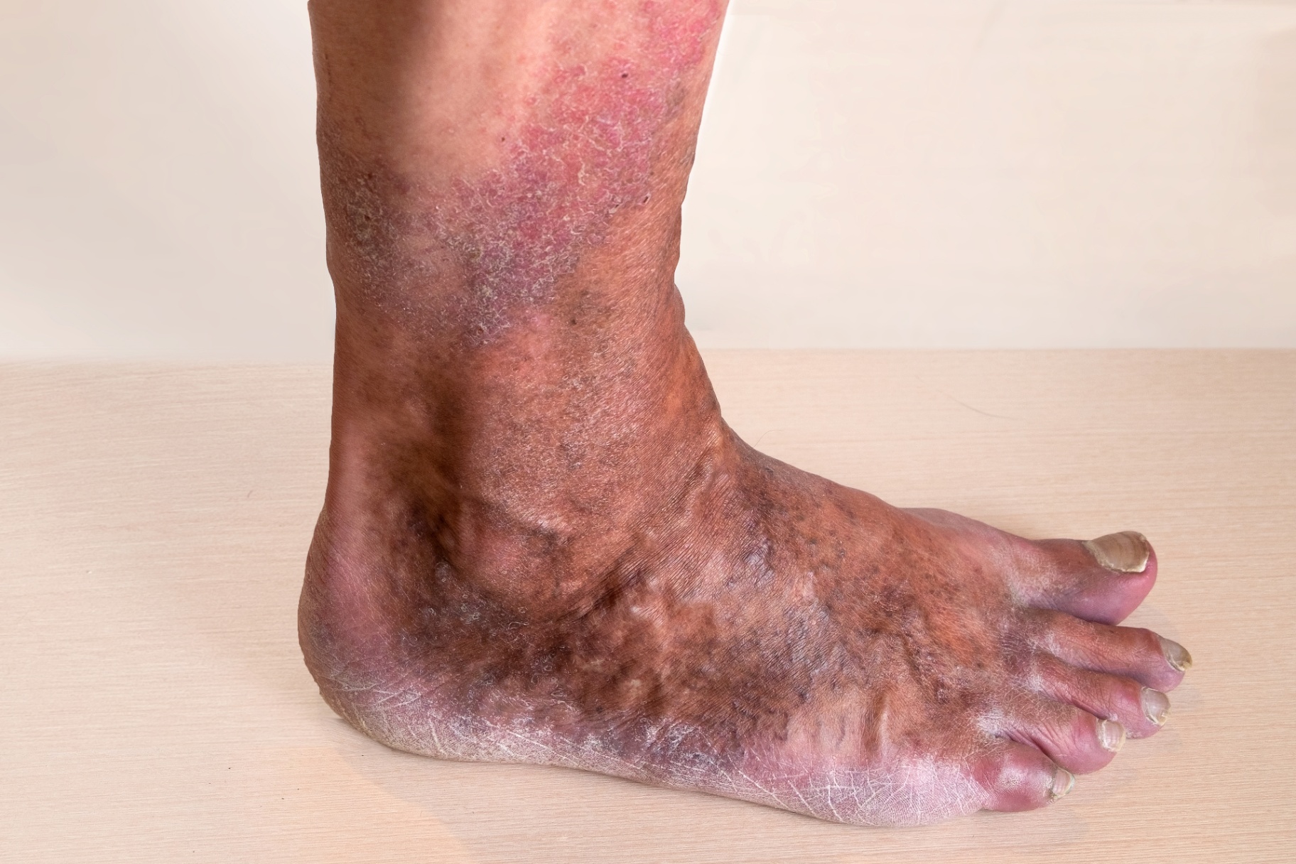
Brown spots, itching and dry, scaly skin on the lower leg are classic signs of venous eczema (stasis dermatitis) - a skin reaction to long-standing venous hypertension. The condition is one of several skin findings that can result from chronic venous insufficiency [1]. It is seen more often with more advanced CEAP-classified venous disease, but on its own it does not predict the course in an individual patient. In the Edinburgh Vein Study, a population survey of adults aged 18-64 in Scotland followed for 13 years, about 9% developed new chronic venous insufficiency, corresponding to an incidence of roughly 0.7% per year, with risk rising with age [2]. That is a cohort estimate from one urban population, not a universal risk. At population level, untreated venous hypertension is associated with a higher occurrence of venous ulcers at higher CEAP classes, but this cannot be used to predict the course for an individual.
How does venous eczema develop?
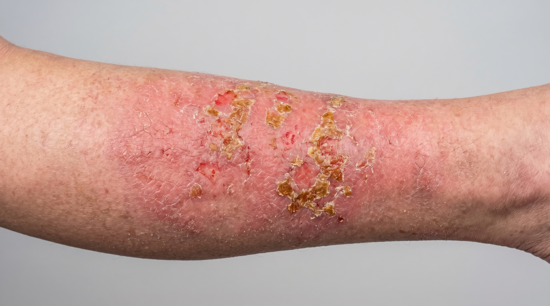
When the venous valves fail, blood flows backwards (reflux) and creates elevated pressure in the veins of the lower leg. This causes:
- Leakage of red blood cells through the capillary wall
- Haemosiderin deposits - the iron in haemoglobin permanently colours the skin brownish
- Chronic inflammation in the subcutaneous tissue
- Oxygen deficiency in the skin → dry, itchy, scaly surface
- Fibrosis (lipodermatosclerosis) → firm, constricted skin around the ankle ("inverted champagne bottle")
Symptoms you should know
| Sign | What it means |
|---|---|
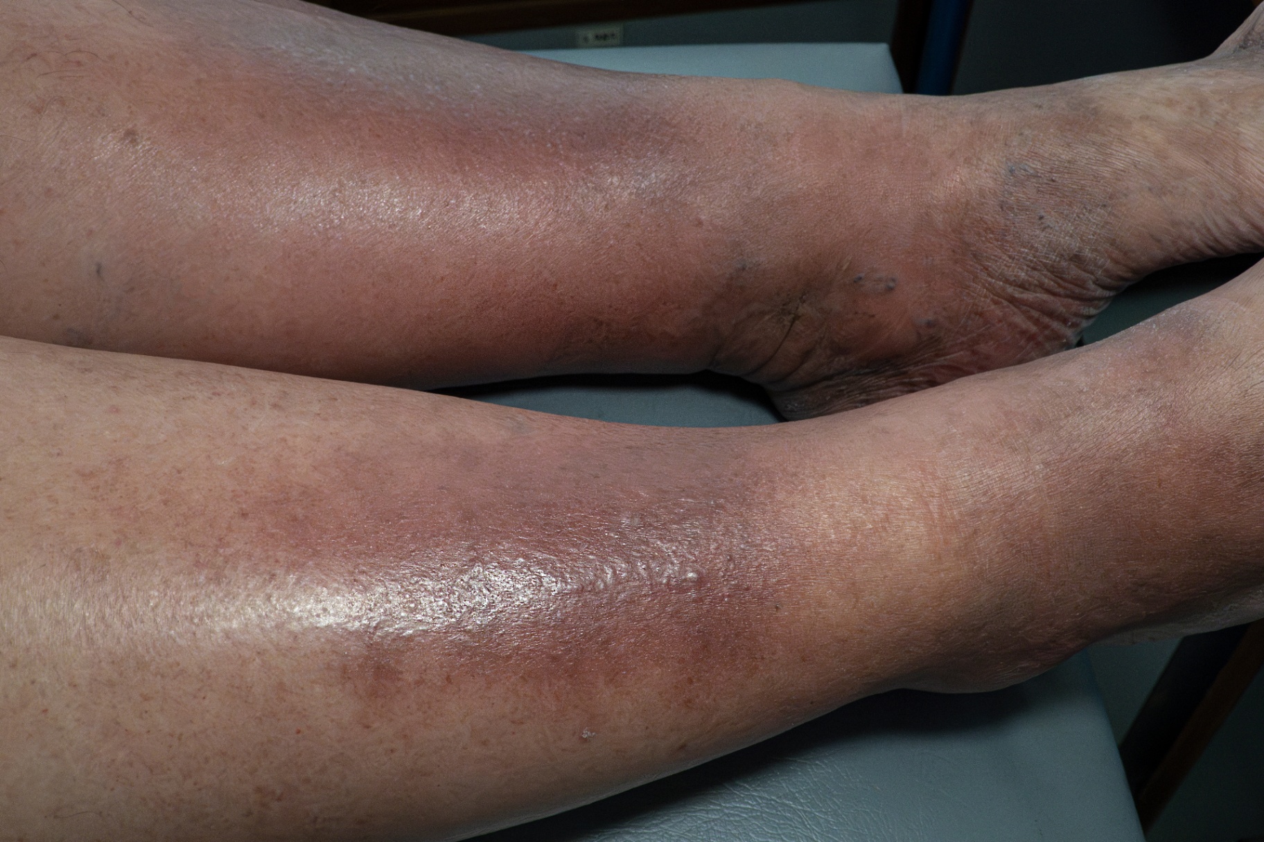
| Brown spots on the inside of the ankle | Haemosiderin - chronic venous hypertension |
| Itching and dry skin on the lower leg | Active venous eczema (C4a) |
| Scaly or weeping skin | Inflammatory phase |
| Hard, constricted skin at the ankle | Lipodermatosclerosis (C4b) - associated with higher ulcer occurrence in study populations |
| White scarred areas | Atrophie blanche (classified as C4c in CEAP 2020) - often seen in patients who have or have had venous ulcers (C5-C6) |
| Swelling at the end of the day | Associated swollen legs |
How to distinguish venous eczema from other skin disease
| Diagnosis | Location | Characteristics |
|---|---|---|
| Venous eczema | Inside of ankle/calf | Brown pigmentation, concurrent varices/oedema |
| Contact eczema | Wherever the allergen touches | Acute, itchy, possibly blisters |
| Atopic eczema | Elbow folds, knees, neck | Personal/family atopy |
| Cellulitis | Acute, unilateral | Fever, warmth, intense redness |
| Arterial ischaemia | Toes, dorsum of foot | Pain, cool skin, weak pulses |
Venous eczema can be mistaken for cellulitis, and a secondary bacterial skin infection can also occur alongside it. Antibiotics do not treat the eczema itself, but may be relevant if an infection is confirmed after medical assessment [3].
When to see a doctor?
Contact your GP or a specialist surgeon if you have:
- Brown spots or discolouration on the lower leg
- Itchy, scaly skin for several weeks
- Visible varicose veins + skin changes
- Hard, firm skin around the ankle
- Previous venous ulcer
- Associated heavy, tired legs or spider veins
Work-up
- Clinical assessment and CEAP classification (C4a/C4b).
- Doppler ultrasound (duplex scan) - maps valve failure and reflux [4].
- Arterial assessment: clinical assessment of arterial status, and ABPI or another measurement when indicated, forms part of the evaluation. The compression level and whether compression is suitable depend on arterial status, symptoms, wound status and the overall clinical picture. With arterial insufficiency, reduced compression or a contraindication may be relevant after specialist assessment.
- Patch test if secondary contact allergy to creams or dressings is suspected.
Treatment
Treatment is planned individually by the treating doctor or nurse based on clinical assessment, arterial status and the severity of the eczema. The following is a general description, not a self-treatment plan.
1. Treating the underlying venous cause:
- Endovenous laser ablation (EVLA/EVLT) of incompetent trunk veins is one option when reflux is documented; the method is chosen individually.
- Treating the reflux addresses the cause of the high pressure in the veins. What that means for the eczema itself has not been settled in randomised trials. The randomised EVRA trial showed that venous ulcers healed faster when early endovenous ablation was combined with compression, but the trial looked at ulcer healing, not eczema [5].
2. Compression:
- Compression stockings can be part of treatment. The compression level chosen, and whether compression is suitable at all, depends on arterial assessment (ABPI), symptoms, wound status and an overall clinical judgement.
- In arterial insufficiency, reduced compression or a contraindication to compression may be relevant, following assessment by a specialist.
3. Topical treatment of acute eczema:
- Moisturiser (unscented) can help with dry skin; how often to apply it is decided individually with the treating doctor or nurse
- Steroid cream may be used for active inflammation; the strength and duration of treatment is a medical or dermatological judgement and should not be decided independently [6]
- Gentle skin care instead of soap and wet wipes may reduce irritation
- Known contact allergens such as lanolin and perfumed products should be avoided if contact allergy is suspected
4. Lifestyle:
- Physical activity can support the calf muscle pump
- Elevating the legs for short periods may ease symptoms
- Weight loss can be considered in overweight individuals, after individual assessment
- Long periods of standing or sitting should be limited where possible
Treatment at Kirurgen.dk
We offer Doppler ultrasound and endovenous laser ablation (EVLA/EVLT) after a GP referral. Questions about referral, cover and price are clarified with the clinic or your GP before treatment; see contact. Whether early treatment of reflux can prevent a venous ulcer is not settled; the randomised EVRA trial studied healing of ulcers that had already developed, not prevention. See also our articles on treatment of varicose veins and causes of varicose veins.
References
- Sundaresan S, Migden MR, Silapunt S. Stasis dermatitis: pathophysiology, evaluation, and management. Am J Clin Dermatol 2017;18(3):383-90.
- Robertson L, Lee AJ, Evans CJ, et al. Incidence of chronic venous disease in the Edinburgh Vein Study. J Vasc Surg Venous Lymphat Disord 2013;1(1):59-67.
- Weaver J, Billings SD. Initial presentation of stasis dermatitis mimicking solitary lesions. J Am Acad Dermatol 2009;61(6):1028-32.
- Gloviczki P, Lawrence PF, Wasan SM, et al. The 2022 SVS/AVF/AVLS clinical practice guidelines for the management of varicose veins of the lower extremities. Part I. Duplex scanning and treatment of superficial truncal reflux. J Vasc Surg Venous Lymphat Disord 2023;11(2):231-261.e6. DOI: 10.1016/j.jvsv.2022.09.004. PubMed
- Gohel MS, Heatley F, Liu X, et al. Early endovenous ablation in venous ulceration (EVRA trial). N Engl J Med 2018;378(22):2105-14.
- Nedorost ST. Generalized dermatitis: a practical clinical approach. Dermatol Ther 2012;25(2):143-8.
More on this topic at Kirurgen.dk
Category: Varicose veins