
Peptic ulcer disease
Written and medically reviewed by Bahir Hadi, specialist in surgery, PhD
Peptic ulcer disease: symptoms, causes, diagnosis and treatment

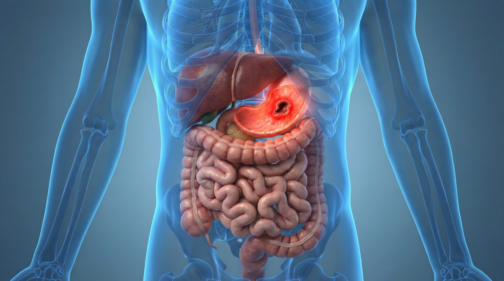
Peptic ulcer disease (PUD) is a defect in the mucosa of the stomach (gastric ulcer) or duodenum (duodenal ulcer) that breaks through the mucosa's own muscle layer (muscularis mucosae) and reaches the underlying tissue. Superficial defects that do not reach that deep are called erosions. The prevalence of peptic ulcer has fallen substantially over recent decades, partly because H. pylori infection has become less common and acid-suppressing treatment is widespread. Management has become more challenging, however, because of antimicrobial resistance and widespread use of antithrombotic drugs in an ageing population [1]. The two dominant causes are Helicobacter pylori infection and use of NSAIDs (ibuprofen, diclofenac) or aspirin, while a smaller group have other or unidentified causes.
Symptoms
The classical symptom is burning or gnawing epigastric pain:
- Duodenal ulcer: pain 2-5 hours after meals and at night - relieved by food or antacids.
- Gastric ulcer: pain triggered or worsened by eating - patients often lose weight.
Other symptoms: nausea, bloating, early satiety, heartburn, acid regurgitation.
Signs of acute bleeding - contact the acute helpline the same day (1813 in the Capital Region of Denmark, otherwise your local out-of-hours service), and call 112 if the circulation is affected:
- Black tarry stool (melena)
- Vomiting blood or coffee-ground material
- Fainting, severe dizziness, pallor or a rapid pulse
Other alarm symptoms - need prompt medical work-up, but not automatically 112:
- Unexplained weight loss
- Persistent vomiting
- Progressive dysphagia
- Iron-deficiency anemia
Causes
| Cause | Mechanism |
|---|---|
| Helicobacter pylori | Disrupts the mucosal barrier. Acid output may be increased (antral inflammation, typical of duodenal ulcer) or reduced (corpus inflammation, typical of gastric ulcer) |
| NSAID/aspirin | Inhibits COX-1 → reduced prostaglandin protection |
| Stress ulcer | ICU patients, burns, critical illness |
| Zollinger-Ellison syndrome | Gastrin-producing tumor → extreme acid output |
| Unexplained | No identifiable trigger |
The doctor's risk assessment takes account of factors such as smoking, alcohol, age and concurrent treatment with corticosteroids or blood thinners.
Diagnosis
- Oral or nasal gastroscopy - the central investigation. Direct visualization and a CLO test for H. pylori. Gastric ulcers are biopsied to exclude malignancy, whereas duodenal ulcers are usually not biopsied. During acute bleeding the procedure belongs in hospital.
- H. pylori testing: the ¹³C-urea breath test or a stool antigen test detects active infection and is also used to confirm eradication. Serology (blood antibodies) can stay positive after an infection has cleared and does not separate active from past infection, so it is not an equivalent alternative. Acid-suppressing drugs and antibiotics can cause a false negative breath or stool test, and any pause is agreed with whoever orders the test.
- Blood tests: hemoglobin, ferritin (iron deficiency from chronic bleeding), gastrin (suspected ZES).
- Repeat gastroscopy for gastric ulcers to confirm healing and rule out cancer. The timing is set individually and is usually some months after treatment starts; ASGE recommends follow-up endoscopy for gastric ulcers, while the exact interval differs between guidelines [2]. Uncomplicated duodenal ulcers are usually not re-scoped, but eradication must be documented.
Gastroscopy makes it possible to look directly at the lining and take tissue samples, but it is not needed for every pain in the upper abdomen. The choice is made after an overall assessment that includes alarm features and Helicobacter pylori status.
Treatment
First-line - H. pylori eradication (positive test):
- The regimen is chosen by the doctor based on local resistance, previous antibiotic exposure and allergies. Clarithromycin-based triple therapy should not be used uncritically as resistance is rising; bismuth quadruple therapy is an alternative.
- Test of cure is mandatory: breath test or stool antigen no earlier than 4 weeks after antibiotics are finished, and after an agreed PPI pause. See the breath test.
- Eradication substantially lowers the risk of a new ulcer compared with acid suppression alone [3].
General ulcer healing:
- Proton pump inhibitor (PPI). Dose and duration are set by the doctor based on the type and cause of the ulcer, together with an agreed point for review.
- NSAIDs, aspirin and blood thinners are reviewed by the doctor, who decides on pausing, switching or adding PPI cover. Never stop prescribed medication on your own.
- Smoking cessation and alcohol reduction accelerate healing.
Surgery (rare - complications only):
- Perforation (acute peritonitis → emergency laparotomy)
- Uncontrolled bleeding despite endoscopy
- Gastric outlet obstruction
- Suspected malignancy
Complications
- Bleeding (the most common complication) - presents as melena or hematemesis.
- Perforation (less common, but acute and serious) - sudden, knife-like abdominal pain, "board-like" abdomen.
- Penetration into pancreas - back pain, raised amylase.
- Stenosis (rare) - vomiting of undigested food.
Prevention
- Test and treat H. pylori in patients with family history of gastric cancer.
- Consider PPI cover with long-term NSAID treatment in older patients or those with previous ulcers, after medical assessment.
- Minimise NSAID use and consider whether paracetamol can cover the pain instead. The choice depends on the type of pain and other conditions and is made with your doctor.
Further assessment and treatment
What treatment involves depends on the cause. If Helicobacter pylori is found, eradication therapy is central, while for NSAID-induced ulcers the doctor assesses whether the causative drug should be changed or stopped, alongside acid-suppressing treatment. Where there are alarm features or unresolved findings, gastroscopy with tissue samples may be relevant, and whether it is done orally or nasally depends on the individual. Read on about GERD and reflux and nasal vs. oral gastroscopy.
References
- Lanas A, Chan FKL. Peptic ulcer disease. Lancet 2017;390(10094):613-24.
- Banerjee S, Cash BD, Dominitz JA, et al. The role of endoscopy in the management of patients with peptic ulcer disease. Gastrointest Endosc 2010;71(4):663-8.
- Ford AC, Gurusamy KS, Delaney B, Forman D, Moayyedi P. Eradication therapy for peptic ulcer disease in Helicobacter pylori-positive people. Cochrane Database Syst Rev 2016;(4):CD003840. PubMed
More on this topic at Kirurgen.dk
Category: Gastrointestinal